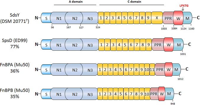
FIG 1.
Schematic representation of SdsY from S. delphini DSM 20771T. The SdsY protein includes a signal sequence (dark blue; S) at the N terminus, followed by an A domain spanning residues 37 to 524 (light blue; N1, N2, N3), a connecting domain in yellow containing nine tandem repeats with an immunoglobulin-like domain X (C; residues 525 to 1003), a proline-rich repeat spanning residues 1004 to 1084 (light red; PPR), and the wall (red; W) and membrane (cyan; M) spanning domains at the extreme C terminus residues 1085 to 1160 (22). The A domain of SdsY has 70% identity with SpsD, 38% identity with FnBPA, and 43% identity with FnBPB. The putative fibronectin binding region of SdsY has 73% identity with SpsD, 43% identity with FnBPA, and 41% identity with FnBPB.

