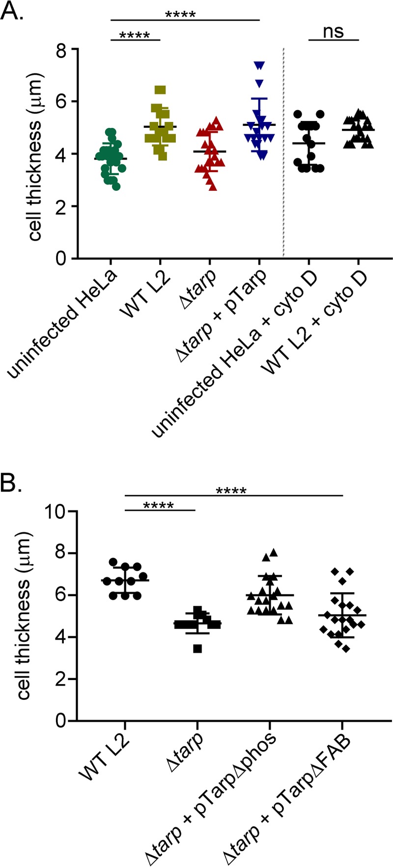
FIG 5.

Chlamydia trachomatis invasion of HeLa cells alters the host cell shape. (A) Wild-type Chlamydia trachomatis L2 (WT L2, light green squares), tarP mutant (ΔtarP, red triangles), or tarP mutant complemented with pTarp (Δtarp + pTarp, blue upside down triangles) underwent chlamydial invasion of HeLa 229 cells for 30 min. Bacteria were visualized by staining with anti-MOMP primary antibody followed by goat anti-mouse Alexa Fluor 488 secondary antibody. The actin cytoskeleton was stained with Alexa Fluor 568 Phalloidin. The cell thickness was compared to uninfected HeLa cells (uninfected HeLa, green circles) by z-stack analysis on a Zeiss 710 inverted confocal microscope. Samples were compared using one-way ANOVA and Tukey’s multiple comparison test. ****, P < 0.0001. The graph presented is from one representative experiment of three performed. Host cells pretreated with the actin-destabilizing drug cytochalasin D (uninfected HeLa + cyto D, black circles) or drug-treated cells infected with wild-type C. trachomatis (WT L2 + cyto D, black triangles) did not undergo changes to cell shape. (B) Similarly, HeLa 229 cells were infected with wild-type Chlamydia trachomatis L2 (WT L2, black circles), tarP mutant (Δtarp, black squares), or tarP mutant complemented with pTarpΔphos (Δtarp + pTarpΔphos, black triangles) or pTarpΔFAB (Δtarp + pTarpΔFAB, black diamonds), and the cell thickness was quantified as described in panel A.
