
FIGURE 1.
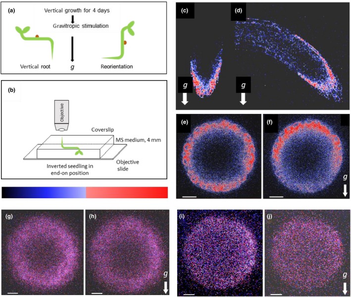
Fluorescence micrographs of vertical and 90° tilted roots expressing EHB1‐GFP, AGD12‐GFP, and GFP under control of a 35S‐CaMV promoter, respectively. (a) Experimental setup for a gravitropic stimulus. Seedlings were grown vertically for 68 hr in the dark, presented in C, E, G, and I, respectively. (b) After that seedlings were tilted 90° and left for additional 101 min in the dark shown in D, F, H, and J. (c,d) Images of EHB1‐GFP fluorescence in which the axis of the microscope objective is perpendicular to the root axis. (e,f) Same seedlings analyzed from the root tip (“end on”). (e) Vertically grown roots revealed a symmetric distribution of EHB1‐GFP. (f) In reoriented roots, EHB1‐GFP fluorescence got asymmetrically distributed or polarized with enhanced fluorescence at the top and reduced fluorescence at the bottom. AGD12‐GFP (g,h) and GFP alone (i,j) expressing roots do not show any gravitropically induced redistribution. Images were taken 80 µm above the root tip. g = direction of the gravitational acceleration. Scale bar = 15 µm. The relative fluorescence in this and the following figures were quantified and displayed as heat map, were high fluorescence is shown in red and moderate expression in blue