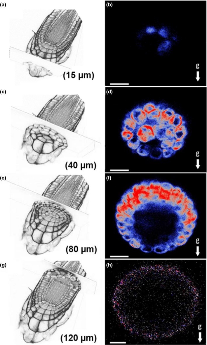
FIGURE 3.

EHB1‐GFP expression in roots that were kept horizontally for 48 min prior to microscopy. Root caps were either scanned at 15 µm (a,b), 40 µm (c,d), 80 µm (e,f), and 120 µm (g,h) depth, respectively. Left: section overview obtained from phase‐contrast microscopy. Right: confocal images of the EHB1‐GFP asymmetry in dependence from the distance to the root tip (end‐on visualization). EHB1 is located preferentially in the peripheral layers of the root cap between 40 and 80 µm, in which also the asymmetry is observed after gravistimulation. EHB1‐GFP is also found at 40 µm in the central statocytes of the columella (d). g = gravitational acceleration. Scale bar = 15 µm
