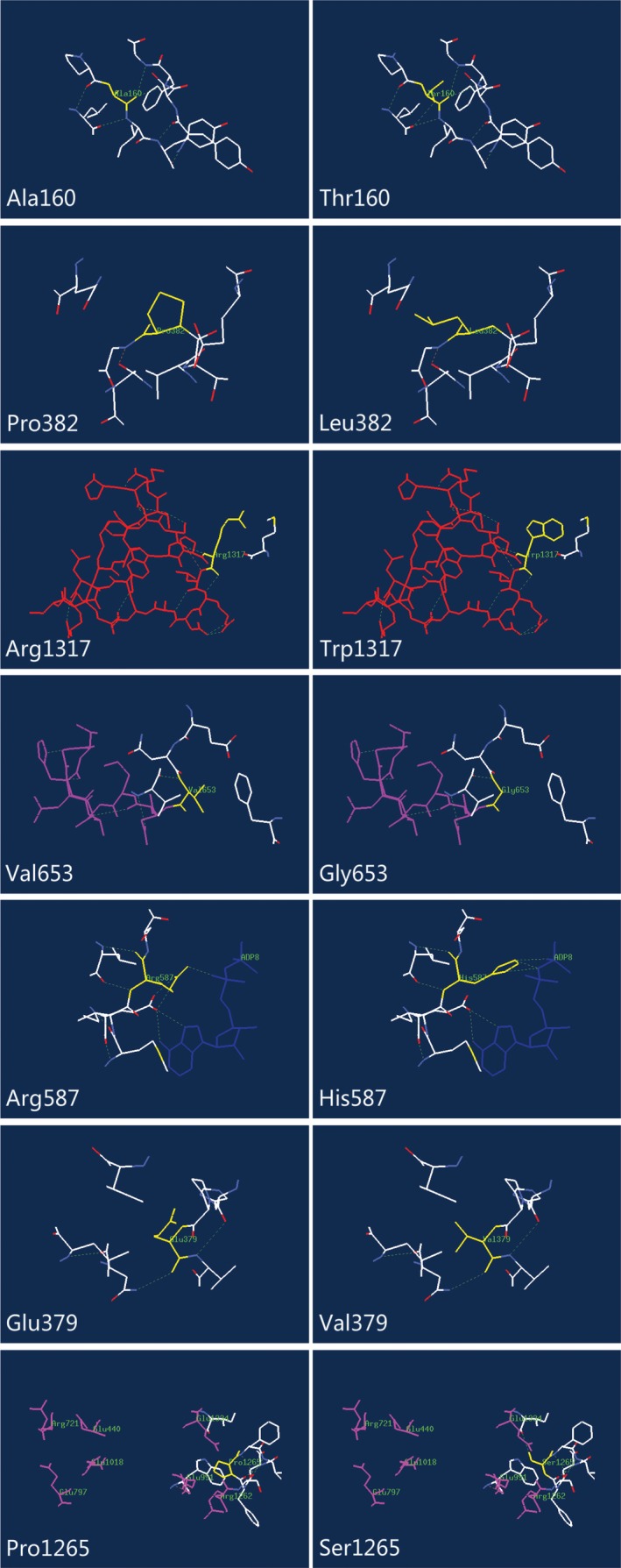
Figure 2.

Structural analysis of wild‐type (WT) and the variant CPS1 with mutations. The residues of missense mutant sites together with the nearby functional site are illustrated in WT and variant CPS1 using Swiss‐Pdb Viewer. The computed hydrogen bonds are shown as green dashed lines. Residues of the mutant sites are highlighted in yellow. T'‐loop (L3β15‐L3β16 loop, residues 1311‐1333) is shown in red, K‐loop (L1β11‐L1β12, residues 654‐662) is shown in purple, molecular ADP is shown in deep blue, and seven amino acids contributional to the postulated carbamate tunnel are highlighted in pink. The nearby residues within 5 Å are shown in white
