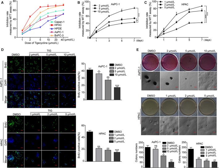
Figure 1.

Tigecycline inhibits cell growth and proliferation in human PDAC cell lines. A, Inhibition rates of the four human PDAC cell lines and one human normal pancreatic duct glandular cell line (HPDE) treated with increasing concentrations of tigecycline for 72 h. B, C, Inhibition rates of AsPC‐1 and HPAC cells after treating with different concentrations of tigecycline for the indicated time by using MTT assay. D, Image and quantification of AsPC‐1 and HPAC cells positive for BrdU staining after treating with DMSO or different concentrations of tigecycline for 72 h. Scale bar = 200 μm. E, Colony formation was examined by soft agar assay (1000 cells/well) in AsPC‐1 and HPAC cells after treating with DMSO or different concentrations of tigecycline for 21‐28 d. Colony numbers were counted. DMSO was used as control. Scale bar = 2 mm. All data were shown as the mean ± SD. Student's t test was carried out. *P < .05; **P < .01, ***P < .001. ns, not significant. P‐value <.05 was considered as statistically significant
