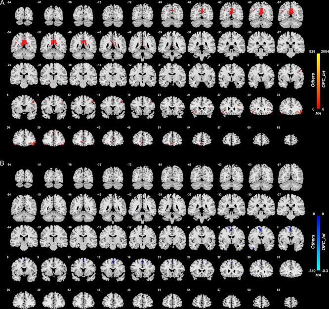
Fig. 1.

Anatomical location of voxels with significantly higher (A) and lower (B) functional connectivity with the lateral orbitofrontal cortex areas in non-medicated depression (patients—controls) obtained from the voxel-based Association Study. Blue indicates voxels with lower functional connectivity in depressed patients, and red/yellow indicates voxels with higher functional connectivity. In this and in all other figures, the level of statistical significance for the difference in functional connectivity for any voxel after correction for multiple comparisons was P < 0.05 FDR.
