
Fig. 2.
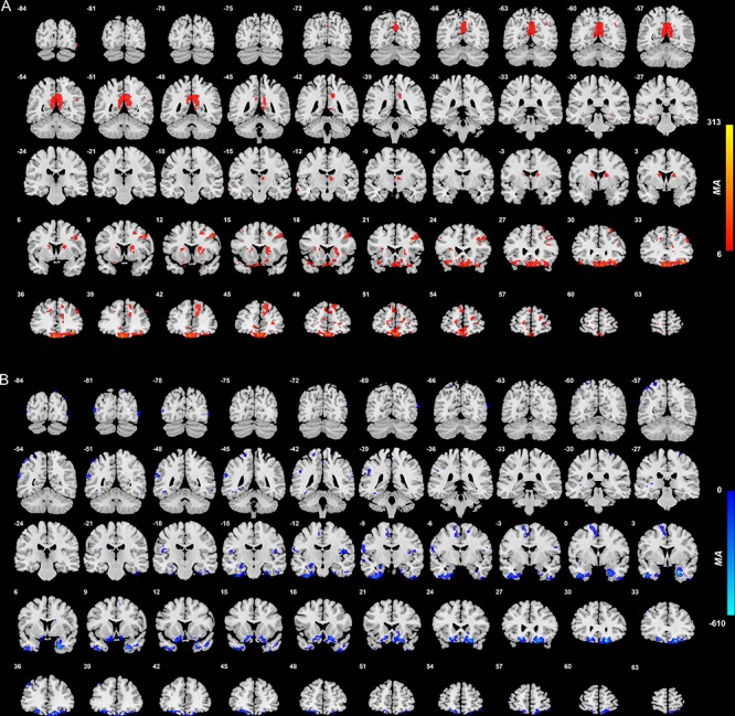
Anatomical location of voxels with significantly increased (A) and decreased (B) functional connectivity with the medial orbitofrontal cortex areas in non-medicated depression (patients—controls) obtained from the voxel-based Association Study. Blue indicates voxels with lower functional connectivity in depressed patients, and red/yellow indicates voxels with higher functional connectivity.