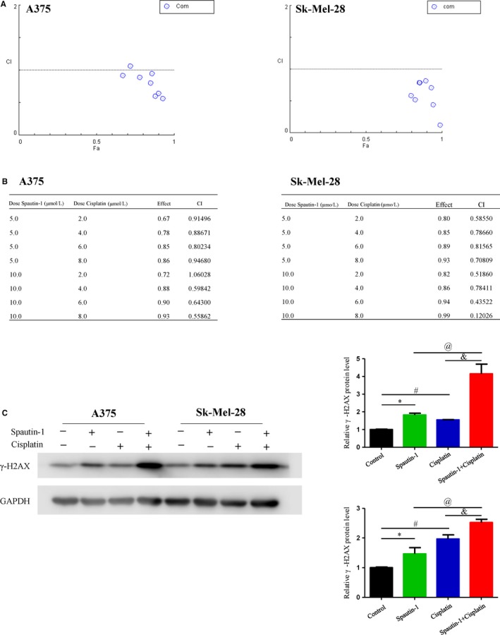
Figure 6.

Spautin‐1 had synergistic effect with cisplatin in the treatment of melanoma cells by induction of DNA damage in vitro. A and B, The synergistic effect of spautin‐1 in combination with cisplatin on the growth of A375 and SK‐Mel‐28 cells. Combination index (CI) values were calculated at the drug concentration of spautin‐1 (5 μmol/L) plus cisplatin (2 μmol/L), spautin‐1 (5 μmol/L) plus cisplatin (4 μmol/L), spautin‐1 (5 μmol/L) plus cisplatin (6 μmol/L), spautin‐1 (5 μmol/L) plus cisplatin (8 μmol/L), spautin‐1 (10 μmol/L) plus cisplatin (2 μmol/L), spautin‐1 (2 μmol/L) plus cisplatin (2 μmol/L), spautin‐1 (10 μmol/L) plus cisplatin (4 μmol/L), spautin‐1 (10 μmol/L) plus cisplatin (6 μmol/L) and spautin‐1 (10 μmol/L) plus cisplatin (8 μmol/L) using the Chou‐Talalay method. C, A375 and SK‐Mel‐28 cell lines were treated with DMSO, spautin‐1 (5 μmol/L) and/or cisplatin (4 μmol/L) for 48 h. Western blot analysis of γ‐H2AX protein expression was performed (Left panel). Semi‐quantitative analysis of γ‐H2AX protein expression compared with GAPDH (right panel). (Mean values ± SEM, n = 3) Significant differences were evaluated using a one‐way ANOVA. *P < .05 Sputin‐1 vs control group, # P < .05 cisplatin vs control group, & P < .05 spautin‐1 vs spautin‐1 + cisplatin, @ P < .05 cisplatin vs spautin‐1 + cisplatin
