
Figure 5.
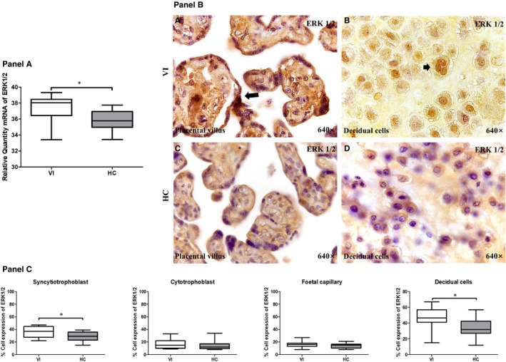
Panel A, ERK 1/2 gene expression between pregnant women with lower extremity venous insufficiency (VI) and pregnant women health control (HC), as measured by RT‐qPCR. Panel B, Histological images of ERK1/2 protein expression showing the relevant immunodetection in the placental villus (C) and decidual cells (D) in the placentas of women with VI. Panel C, Quantification of the percentages of syncytiotrophoblast, cytotrophoblast, foetal capillary and decidual cells that stain positive for ERK 1/2. The arrows are the brown coloration indicating the specific protein expression, and 640× is the magnification of histological images. Data shown median and IQR. *P‐value < .05 (Mann‐Whitney U test)