Figure 2.

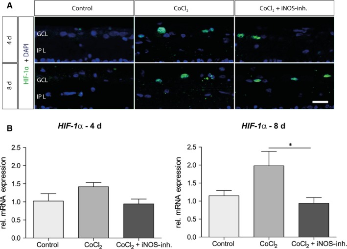
iNOS‐inhibitor mediated effect on hypoxia‐induced HIF‐1α expression. A, Representative pictures of HIF‐1α staining in the ganglion cell layer. HIF‐1α+ cells are marked in green and cell nuclei in blue. The addition of CoCl2 induced an increase or stabilization of the alpha subunit of the transcription factor. Under treatment with the iNOS inhibitor, the HIF‐1a levels were not lower compared with the CoCl2 group after 4 d. In addition, at 8 d of cultivation, comparable amounts of HIF‐1α were detected in CoCl2 and in the 1400W‐treated group. B, After 4 d, no significant differences in HIF‐1α mRNA expression were observed. At 8 d, the relative mRNA expression by CoCl2 was twofold increased and could be lowered to the initial level by treatment with 1400W. Abbreviations: GCL, ganglion cell layer; IPL, inner plexiform layer. Scale bar = 20 µm. All data are shown as mean ± SEM; *P < .05
