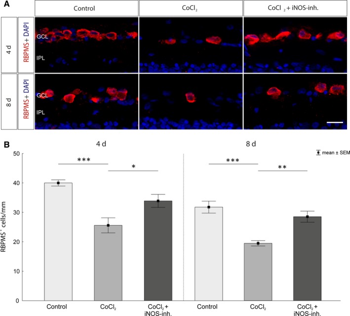
Figure 4.

Rescue of retinal ganglion cells after CoCl2‐induced degeneration. A, Representative images of the immunohistological staining. RGCs were stained with an antibody against RBPMS (red) and cell nuclei with DAPI (blue). A significant loss of RGCs in the untreated degeneration groups (CoCl2) was observed over the cultivation period of 4 and 8 d. B, After 4 d, neuroprotection of the RGCs was observed by treatment with the iNOS‐inhibitor compared with the CoCl2 group. Even after 8 d of cultivation, a protection of the RGCs by 1400W could be noticed. The retinae of the treatment groups contained significantly more RGCs than the untreated retinae. GCL, ganglion cell layer; IPL, inner plexiform layer. Scale bar = 20 µm. All data are shown as mean ± SEM; *P < .05; **P < .01; ***P < .001
