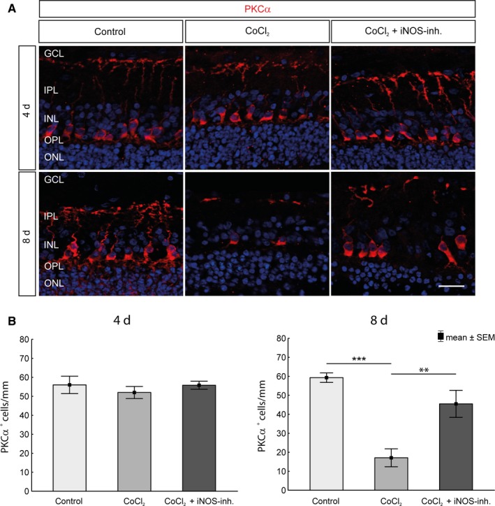
Figure 7.

Protection of bipolar cells against CoCl2‐induced degeneration. A, Nuclei were visualized with DAPI staining (blue), and bipolar cells were stained with an antibody against PKCα (red). B, No significant loss of bipolar cells in the untreated degeneration groups could be detected during the cultivation period of 4 d. After 8 d, a significant loss was observed. However, protection of the bipolar cells by 1400W was observed. The retinae of the treatment group contained significantly more bipolar cells than the untreated retinae. GCL, ganglion cell layer; INL, inner nuclear layer; IPL, inner plexiform layer; ONL, outer nuclear layer; OPL, outer plexiform layer. Scale bar = 20 µm. All data are shown as mean ± SEM; **P < .01; ***P < .001
