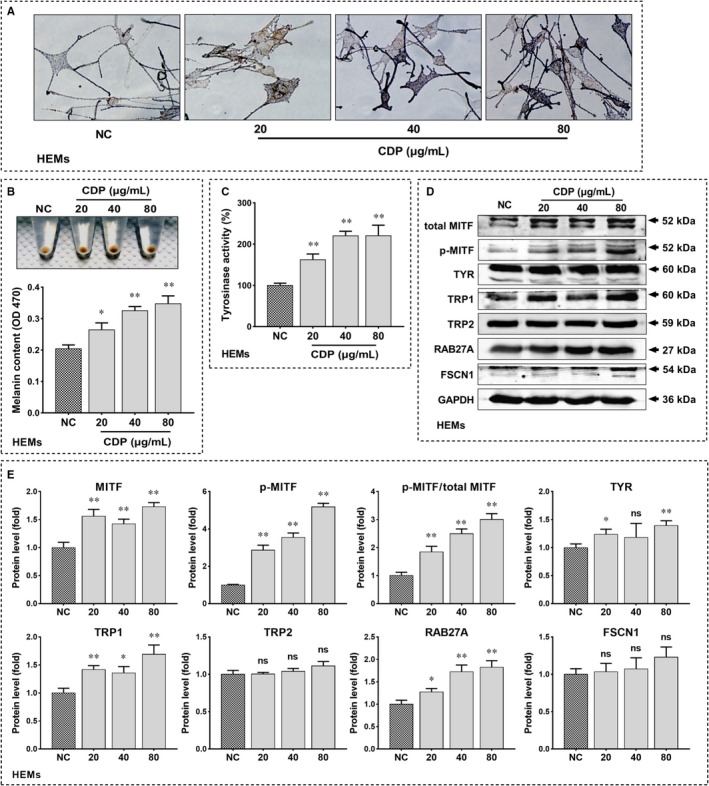
Figure 2.

CDP promotes melanogenesis in HEMs. The HEMs were treated with CDP at different concentrations (20, 40 and 80 μg/mL) or medium alone (NC) for 48 h, then we observed the melanin by Fontana‐Masson staining, measured the melanin content (OD value, 470 nm) by NaOH assay, and measured the tyrosinase activity (OD value, 475 nm) by tyrosinase activity assay; meanwhile, we measured the protein levels of MITF, p‐MITF, TYR, TRP1, TRP2, RAB27A and FSCN1 by Western blotting and measure the grey value of protein bands by Image J: (A) melanin staining; (B) the melanin content; (C) the tyrosinase activity; (D) the protein levels of melanogenesis‐related genes; (E) the statistics of protein's grey value (standardized with GAPDH). (*P < .05, **P < .01)
