Figure 7.

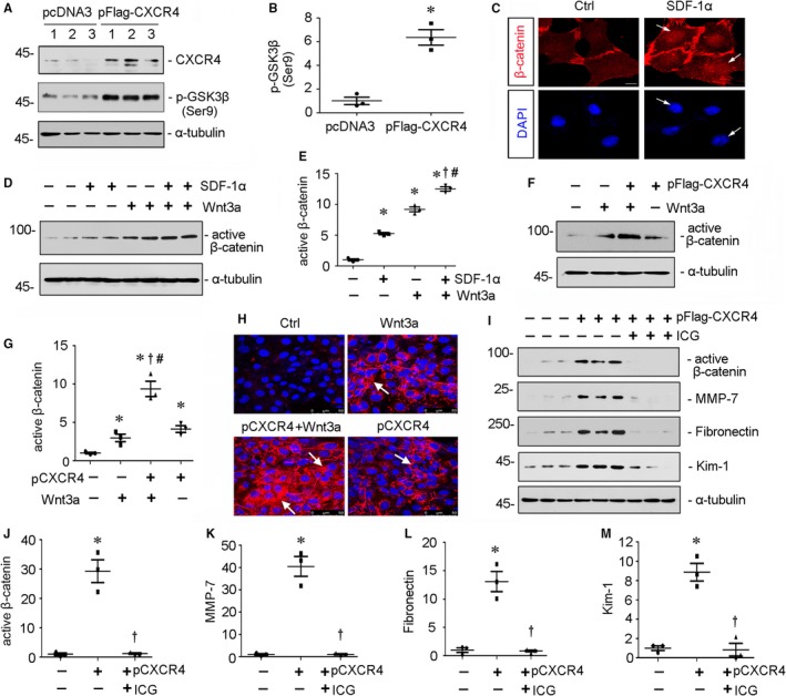
CXCR4/β‐catenin axis plays a critical role in tubular cell injury. A and B, Representative (A) Western blots and graphical representation of (B) p‐GSK3β (Ser9) protein expression in HKC‐8 cells. HKC‐8 cells were transfected with CXCR4 expression plasmid (pFlag‐CXCR4) or empty vector (pcDNA3) for 24 h. Whole cell lysates were analysed by Western blot analyses. Numbers 1‐3 represent each individual well in a given group. *P < .05 versus pcDNA3 group (n = 3). C, Representative micrographs show administration of SDF‐1α induced nuclear translocation of β‐catenin. HKC‐8 cells were treated with SDF‐1α (100 ng/mL) for 12 h. Cells were then stained for β‐catenin and DAPI, the nuclear marker. White arrows indicate positive nuclear staining. Scale bar, 10µm. D and E, Representative (D) Western blots and graphical representation of (E) active β‐catenin protein expression in different groups. HKC‐8 cells were treated with SDF‐1α (100 ng/mL) and/or Wnt3a (100 ng/mL) for 12 h. *P < .05 versus control group (n = 3); †P < .05 versus Wnt3a alone (n = 3); #P < .05 versus SDF‐1α alone (n = 3). F and G, Representative (F) Western blots and graphical representation of (G) active β‐catenin protein expression in different groups. HKC‐8 cells were transfected with pFlag‐CXCR4 or pcDNA3 and then treated with or without Wnt3a (100 ng/mL) for 24 h. *P < .05 versus pcDNA3 group (n = 3); †P < .05 versus Wnt3a alone (n = 3); #P < .05 versus pCXCR4 (pFlag‐CXCR4) alone (n = 3). H, Representative micrographs show fibronectin expression (red) in four groups. HKC‐8 cells were transfected with pFlag‐CXCR4 or pcDNA3 plasmid, then treated with Wnt3a (100 ng/mL) for 24 h. Cells were then immunostained for fibronectin and counterstained with DAPI. White arrows indicate positive staining. I‐M, Representative (I) Western blots and graphical representations of (J) active β‐catenin, (K) MMP‐7, (L) fibronectin and (M) Kim‐1 in three groups. HKC‐8 cells were pretreated with ICG‐001 (5 µM) and then transfected with pFlag‐CXCR4 or pcDNA3 plasmid for 24 h. *P < .05 versus pcDNA3 group (n = 3); †P < .05 versus pCXCR4 (pFlag‐CXCR4) alone (n = 3)
