
Figure 2.
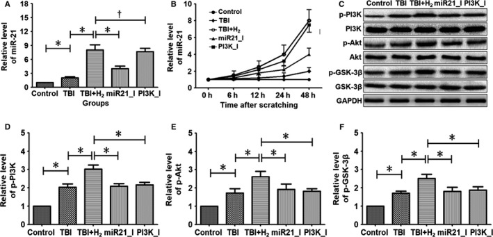
Expressions of miR‐21 and several proteins in PC12 cells. Prepared PC12 cells were randomly divided into control group, TBI group, hydrogen intervention group (TBI + H2 group), miR‐21 inhibition group (miR21_I group) and PI3K inhibition group (PI3K_I group). Each group included 10 wells of PC12 cells. Before scratching, miR‐21 antagomir was transfected into PC12 cells in the miR21_I group, and PC12 cells in the PI3K_I group were treated with PI3K blocker LY294002. After scratching, the injured cells in the TBI + H2 group, miR21_I group and PI3K_I group were cultured in hydrogen‐rich DMEM for 48 h. Expression levels of miR‐21, p‐PI3K, p‐Akt and p‐GSK‐3β were measured using reverse transcription–quantitative PCR, Western blot analysis. Quantitative results were expressed as mean ± standard deviation. Differences in these markers among the five groups were compared using one‐way ANOVA with LSD t test. ‘*’ indicates P < .05 and ‘†’ indicates P > .05