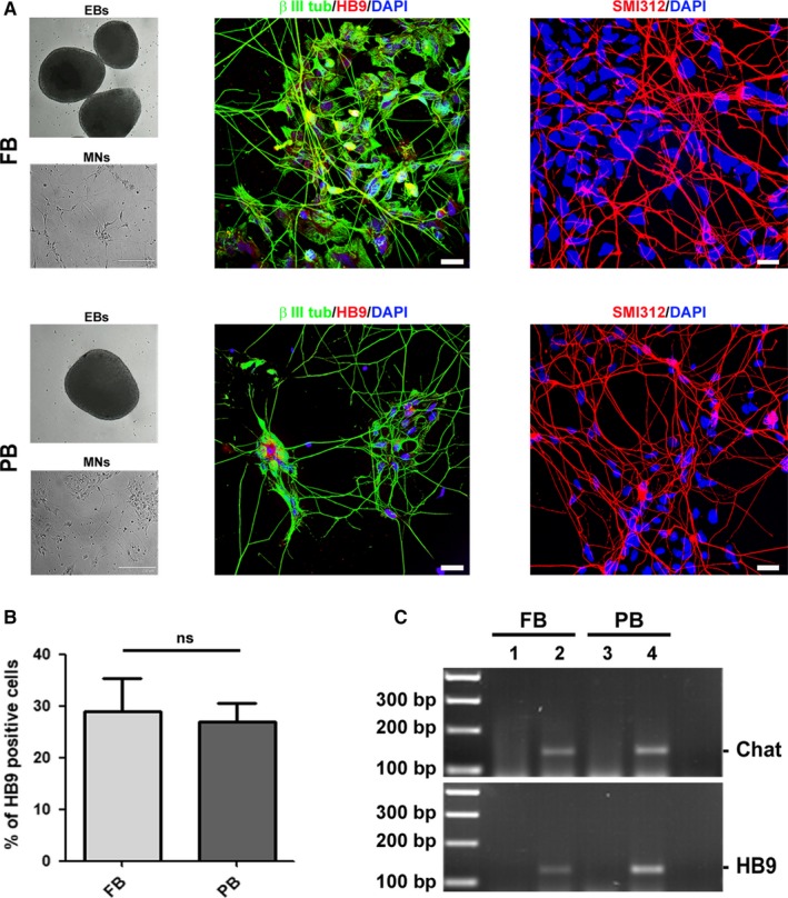
Figure 2.

Comparison of C9ORF72‐mutated iPSC‐derived motor neurons. A, iPSCs from FB and PB were induced to generate embryoid bodies (EBs). After this first step, EBs were dissociated and differentiated into motor neurons (MNs). MNs were stained with the specific nuclear HB9 antigen (red) and co‐stained with the microtubule‐specific marker β III tubulin (green). The positivity for the pan‐axonal neurofilament marker SMI312 was also assessed (red). Nuclei were counterstained with DAPI. Scale bar, 20 µm. B, Histogram displaying the percentage of HB9 positive cells in iPSC‐derived MNs from FB and PB. C, Expression of MNs‐specific markers Chat and HB9 evaluated by RT‐PCR on both iPSCs (lanes 1 and 3) and iPSC‐derived MNs (lanes 2 and 4). Images are representative of 1‐2 independent experiments
