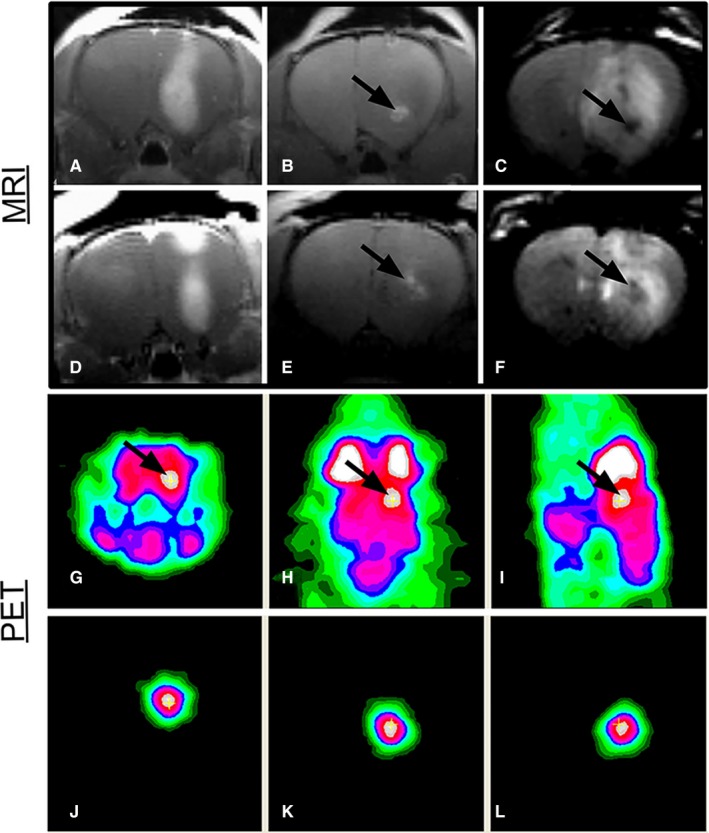
Figure 8.

Gadofullerenes developed for multimodal imaging detection of brain tumours. (A, D) T1‐weighted images with gadodiamide contrast. (B, E) T1‐weighted images in which the bright contrast at the infusion site results from 124I‐f‐Gd3N@C80. (C, F) T2‐weighted images with dark contrast due to 124I‐f‐Gd3N@C80. (G) Coronal, (H) axial and (I) sagittal microPET images following 18F‐FDG injection and the additive image signal that allows for localization of 124I‐f‐Gd3N@C80 within the right hemisphere of the rat brain (arrows indicate infusion sites). (J‐L) MicroPET images display the signal from 124I‐f‐Gd3N@C80. Reprinted with permission from,62 copyright 2012 MDPI (http://creativecommons.org/licenses/by/4.0/)
