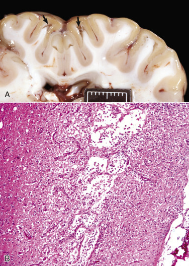
Figure 1-19.

Liquefactive Necrosis.
A, Acute polioencephalomalacia, brain, goat. A thiamine deficiency has resulted in cerebrocortical malacia, which microscopically is liquefactive necrosis with focal tissue separation (arrows). Note yellow discoloration of affected cortex. Scale bar = 2 cm. B, Cortical necrosis, cerebrum, dog. The pale zone in deep laminae of the cerebral cortex is an area of liquefactive necrosis with loss of parenchyma. All that remains is the vasculature with gitter cells in intervening spaces. H&E stain.
(A courtesy Dr. R. Storts, College of Veterinary Medicine, Texas A&M University. B courtesy Dr. L.H. Arp.)
