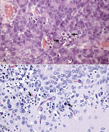
Figure 1-23.

Apoptosis, Cytologic Features.
A, Pancreas, rat. Individual acinar cells are shrunken, condensed, and fragmented (arrows). Apoptotic bodies are in adjacent cells, but inflammation is absent. H&E stain. B, Hippocampus, brain, mouse. Individual neurons are shrunken, condensed, and fragmented (arrows). H&E stain.
(A courtesy Dr. M.A. Wallig, College of Veterinary Medicine, University of Illinois. B courtesy Drs. V.E. Valli and J.F. Zachary, College of Veterinary Medicine, University of Illinois.)
