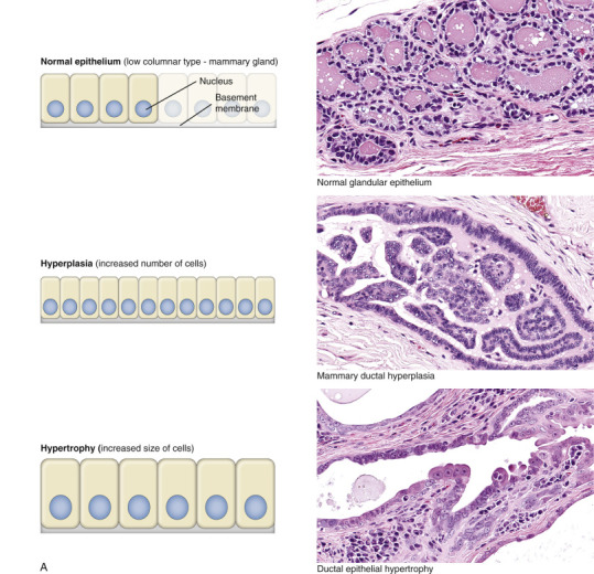
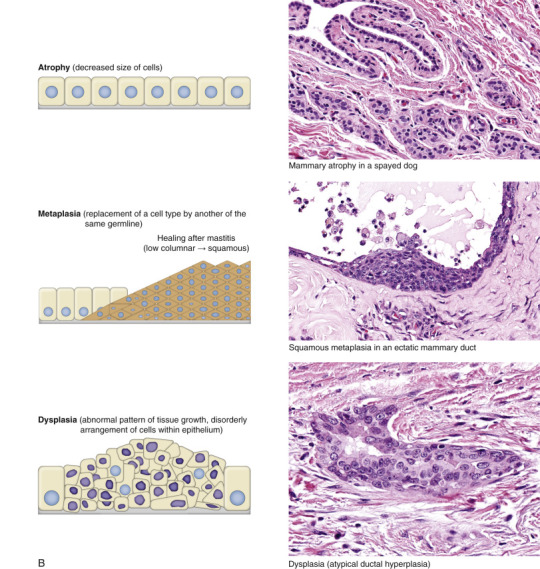
Figure 1-25.


Adaptive Changes Illustrated in Canine Mammary Epithelium.
Schematic diagrams of epithelial adaptations paired with histologic examples from canine mammary glands. A, Normal epithelium, hyperplasia, and hypertrophy. B, Atrophy, metaplasia, and dysplasia. H&E stain.
(Courtesy Dr. M.A. Miller, College of Veterinary Medicine, Purdue University; and Dr. J.F. Zachary, College of Veterinary Medicine, University of Illinois.)
