
Figure 1-32.
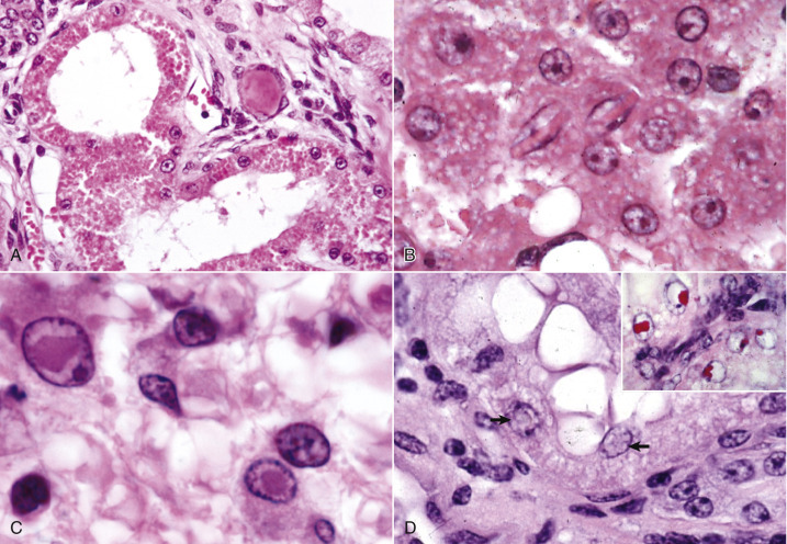
Cell Droplets and Inclusion Bodies.
A, Protein resorption droplets, kidney, dog. The cytoplasm of proximal tubular epithelial cells is filled with eosinophilic droplets—protein that has been resorbed by the cells from the glomerular filtrate. H&E stain. B, Crystalloids, hepatocytes, dog. Note the elongated eosinophilic crystalline inclusions in the nucleus of two hepatocytes. C, Viral inclusion bodies, canine distemper, brain, dog. Note the intranuclear eosinophilic inclusion bodies in astrocytes. H&E stain. D, Lead inclusion bodies, kidney, dog. The intranuclear inclusions (arrows) in renal tubular epithelial cells are difficult to see with an H&E stain. Inset, The lead inclusion bodies are acid-fast (red) and easily observed with Ziehl-Neelsen stain.
(A and C courtesy Dr. M.D. McGavin, College of Veterinary Medicine, University of Tennessee. B courtesy Dr. D.D. Harrington, College of Veterinary Medicine, Purdue University; and Noah's Arkive, College of Veterinary Medicine, The University of Georgia. D courtesy Dr. W. Crowell, College of Veterinary Medicine, The University of Georgia; and Noah's Arkive, College of Veterinary Medicine, The University of Georgia. Inset courtesy Dr. W. Crowell, College of Veterinary Medicine, The University of Georgia; and Noah's Arkive, College of Veterinary Medicine, The University of Georgia.)