E-Figure 1-16.

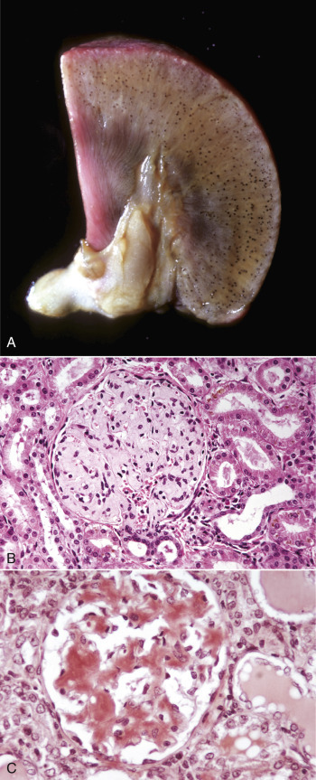
Amyloidosis, Kidney, Dog.
A, Note the blue-black foci, which are glomeruli containing amyloid stained by Lugol's iodine. B, The renal glomerulus contains extracellular deposits of pale homogeneous eosinophilic amyloid. H&E stain. C, The amyloid in the glomerulus stains red-orange. Congo red stain.
(Courtesy Dr. M.D. McGavin, College of Veterinary Medicine, University of Tennessee.)
