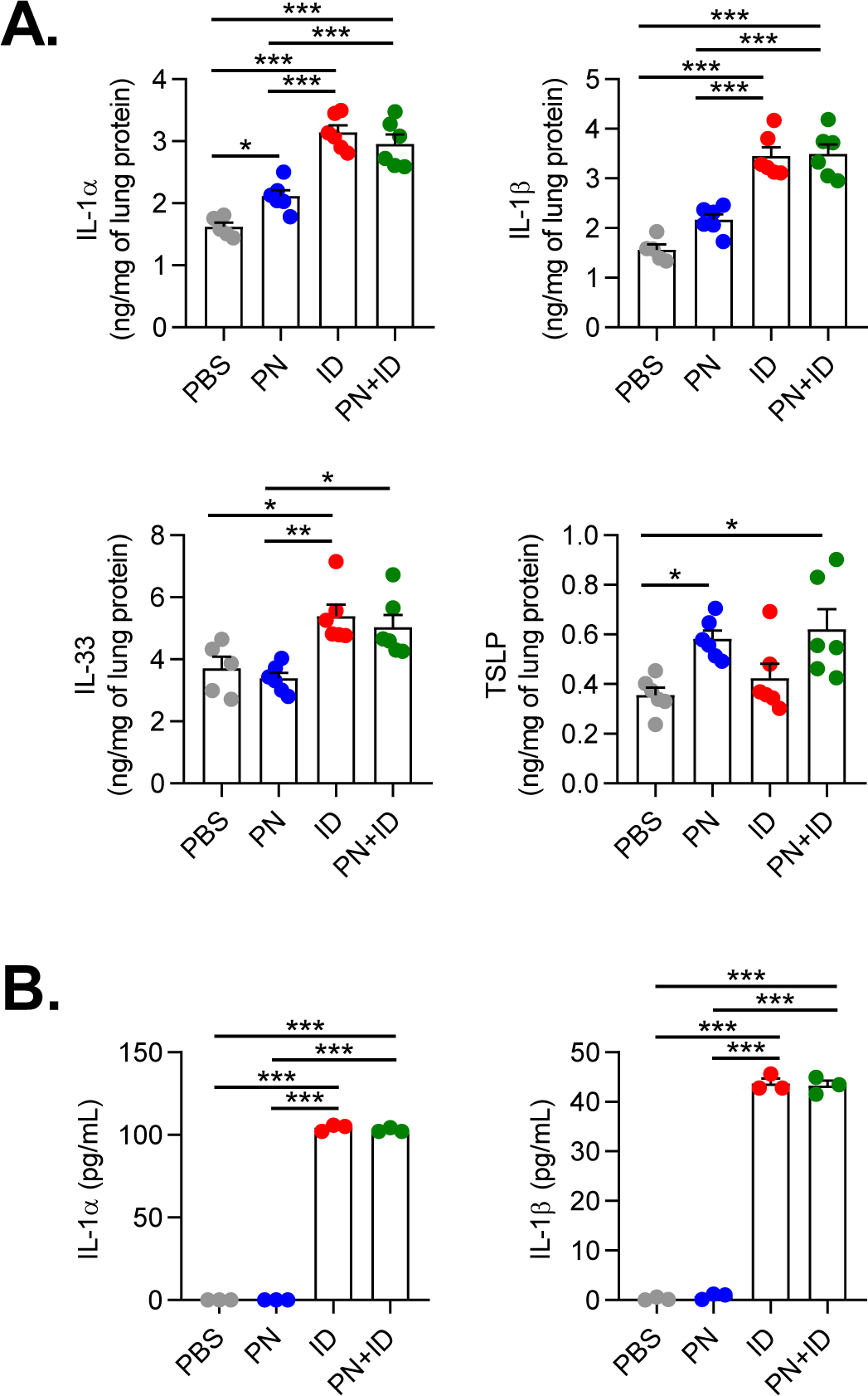
Figure 3. ID induces innate cytokine production in murine lungs and human bronchial epithelial cells.

A. Quantities of IL-1α, IL-1β, IL-33 and TSLP in mouse lung homogenates at 6 hours following exposure to inhaled PBS, PN, ID or PN+ID. Bars represent mean ± SEM, and individual data points are shown (n=5–6 mice per group). B. Quantities of IL-1α and IL-1β in apical cell washes from primary human bronchial epithelial cells at 4 hours following treatment with PBS, PN, ID or PN+ID. Bars represent mean ± SEM, and individual data points are shown (n=3 donors). *P<0.05, **P<0.01, ***P<0.001, one-way ANOVA. PN, peanut; ID, indoor dust.
