
Figure 4. Indoor dust enhances migration of peanut-laden cDC1s to lung-draining LNs.
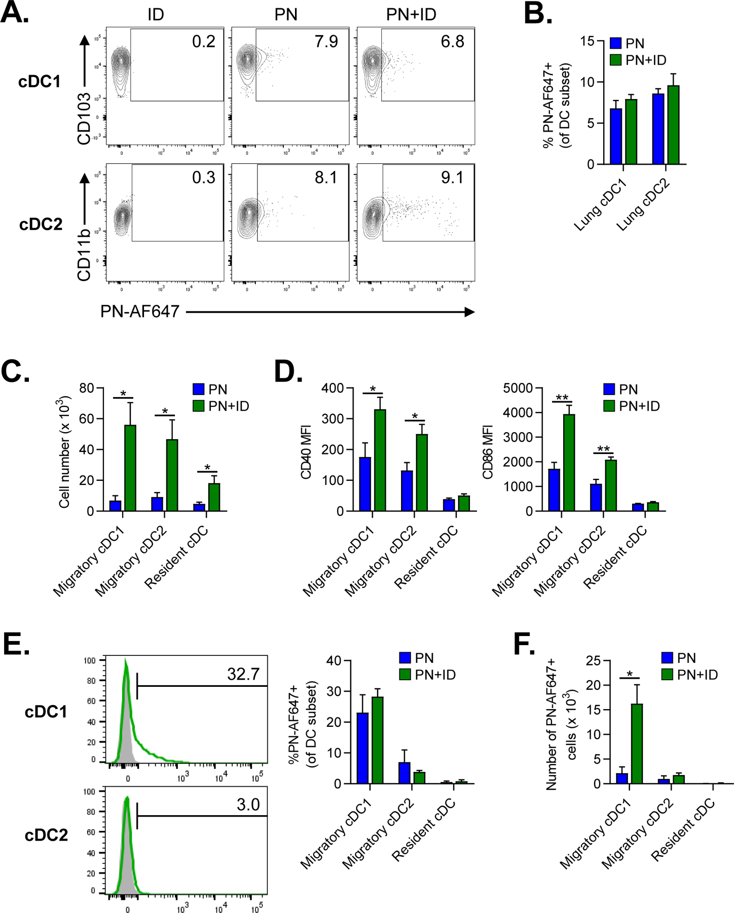
A-B. Flow cytometric analysis of lung cDC subsets 24 hours after airway fluorescent PN-AF647 antigen alone or combined with ID. A. Representative cytograms of PN-AF647 uptake by lung cDC1 or cDC2 subsets. Mice treated with ID alone were included for gating controls. Numbers represent the frequency of cells within each gate. B. Percentage of PN-AF647+ cDC1s and cDC2s in lungs. C-F. Flow cytometric analysis of migratory and resident cDC subsets in lung-draining mLNs at 24 hours following airway instillation of PN±ID. C. Total number of migratory and resident cDC subsets in LNs. D. Median fluorescence intensity (MFI) of CD40 and CD86 expression by cDC subsets in LNs. E. Representative histograms (left) of PN-AF647 uptake by migratory cDC1s or cDC2s in LNs (green histograms). Mice receiving ID alone were included as gating controls (gray histograms). The percentage of PN-AF647+ cDC subsets in LNs is displayed in the graph (right). F. Total number of PN-AF647+ migratory and resident cDC subsets in LNs. Bars represent means ± SEM (n=4–5 mice per group). Data shown are from a single experiment, representative of two experiments. *P<0.05, **P<0.01, Student’s t test. PN, peanut; ID, indoor dust.