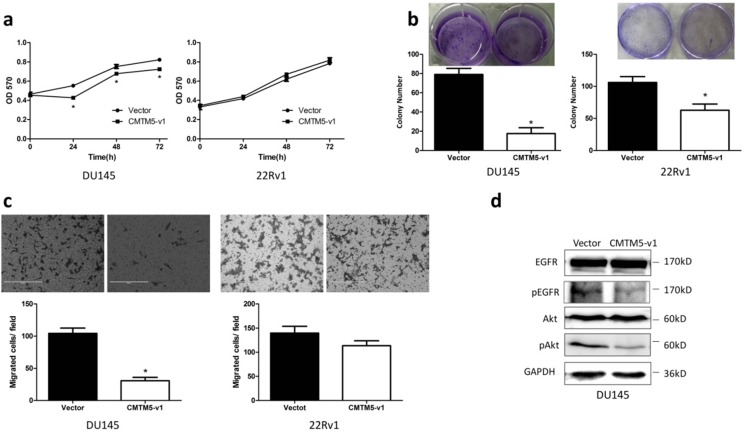
Figure 2.
Effects of CMTM5-v1 on the proliferation and migration of DU145 and 22Rv1 cells. (a) Twenty-four hours after transfection, cells were plated into 96-well plates and then cultured in normal growth medium. At indicated times, cell proliferation was observed using the MTT assay. The results are expressed as the means ± SEM of three independent experiments. (b) Fifteen days after G418 selection, the effect of CMTM5-v1 on colony-forming capacity was measured by counting the number of colonies ≥50 cells. Bars represent the means ± SEM of three independent experiments (*P<0.05). (c) The metastatic potential was determined using a transwell migration assay with medium plus 10% FBS in the bottom chambers. The graph indicates the means ± SEM of the number of cells per three random fields (magnification, x200) counted from three independent experiments (*P<0.05). (d) Forty-eight hours after transfection, DU145 cells were lysed and used to detect the indicated proteins by western blot.

