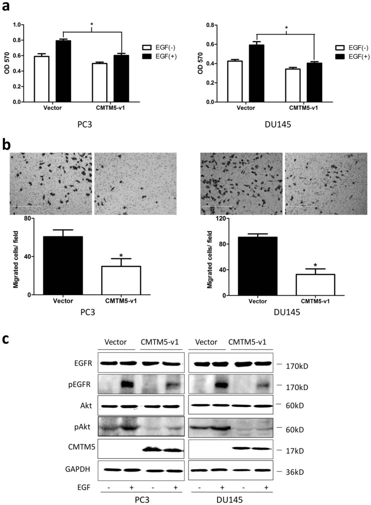
Figure 3.
Effects of CMTM5-v1 on EGF-induced cell growth, migration and EGF-triggered signaling in PC3 and DU145 cells. (a) Twenty-four hours after transfection, cells were plated into 96-well plates and cultured in serum-free medium overnight. The medium was then switched to RPMI-1640 containing 1% FBS in the presence or absence of 20 ng/ml EGF for 48 h. The MTT assay was performed to analyze cell proliferation. Data represent the means ± SEM of the OD570 values of three independent experiments (*P<0.05). (b) The cell migration capacity under EGF-induced chemotaxis was detected in a transwell assay with serum-free medium plus 20 ng/ml EGF in the bottom chambers. Data represent the means ± SEM of cells from three random fields under the microscope, and the experiment was repeated three times. (*P<0.05. Magnification ×200). (c) Transfected cells were serum-starved overnight and then treated with EGF (20 ng/ml) for 5 minutes, and whole-cell lysates were immunoblotted with the indicated antibodies.

