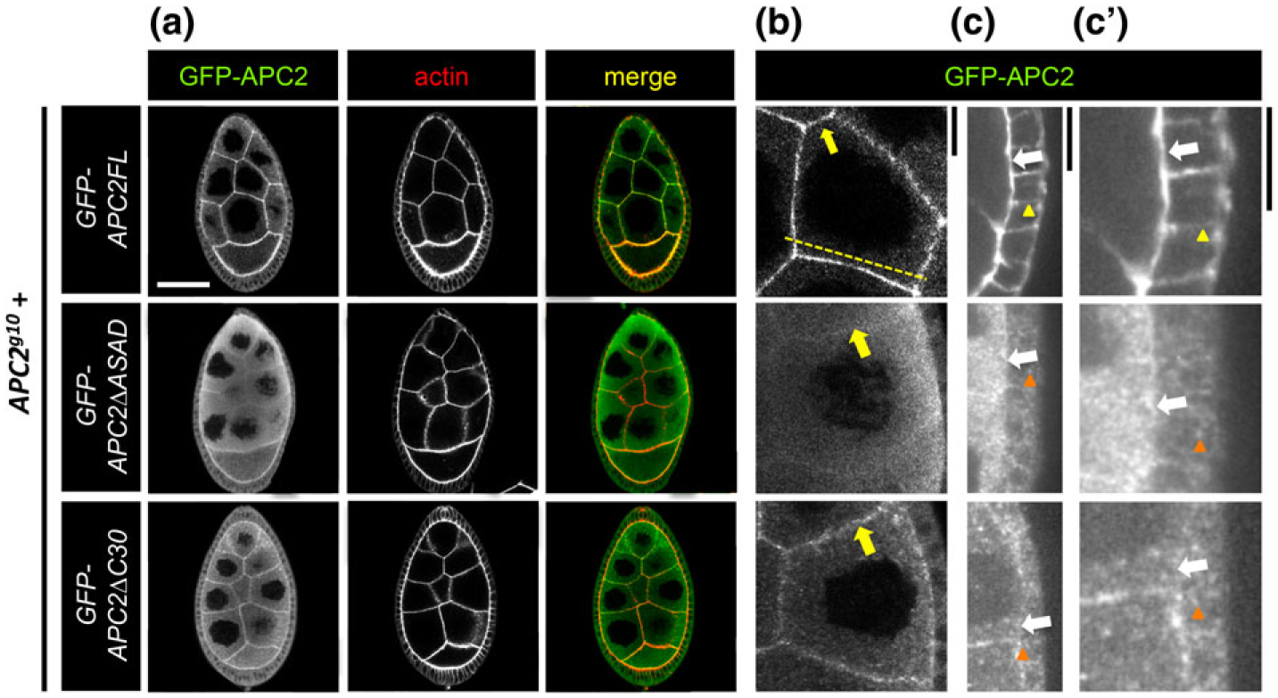
FIGURE 2.

APC2 localization to the nurse cell and follicle cell cortices depends on ASAD and C30. (a-c’) Localization of GFP-tagged APC2FL, APC2ΔASAD, and APC2ΔC30 in the NCs and FCs of the Drosophila egg chamber detected in fixed tissue with anti-GFP. At higher magnification, we saw that APC2FL is cortically enriched in both NCs (b, yellow arrow), and FCs (c,c’, white arrows). APC2FL appears enriched at the apical cortex in FCs (c,c’ white arrow), compared to the basolateral cortex (c,c’ yellow arrowhead), and appears uniform at the NC cortex (b, yellow arrow). Both mutant proteins accumulate significantly in the cytoplasm of NCs and FCs as puncta (a–c’, orange arrowheads). Neither mutant retains significant cortical localization in the FCs (c,c’ white arrows). While the extent of cortical enrichment of the mutant proteins at the NC cortex was variable, APC2ΔASAD tended to have less cortical enrichment than APC2ΔC30 (b, yellow arrows). The dashed line in (b) illustrates the position of a representative line scan shown in Supporting Information Figure S1. Scale bars: (a) 40 μm; (b,c’) 10 μm
