Figure 2.
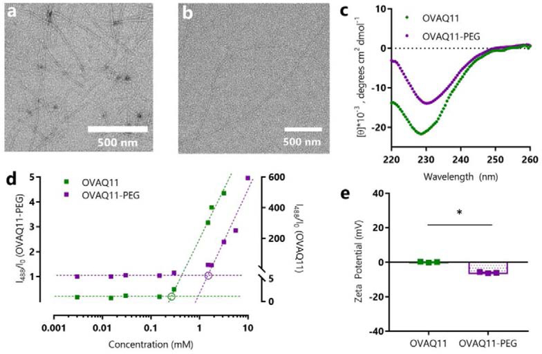
OVAQ11-PEG self-assembled into β-sheet nanofibers, as indicated by negative stained TEM images of (a) OVAQ11 and (b) OVAQ11-PEG nanofibers assembled from 2 mM peptide and (c) Circular dichroism of peptides assembled at 3 mM in PBS and diluted to 0.1 mM in potassium fluoride immediately prior to analyzing. (d) β-sheet structure was further confirmed using Thioflavin T. Following the method of Hamley and coworkers,56 the graphical estimates of critical aggregation concentration correspond to the intersection of the pre- and post-assembly tangent lines (circled). (e) Zeta-potentials of OVAQ11-PEG and OVAQ11 indicated that surface charge was minimally altered by PEGylation. Peptides were prepared at 2 mM in 1X PBS and diluted to 0.2 mM in 1X PBS prior to measurement at 25 °C. * p < 0.05, unpaired, two-tailed T-test

