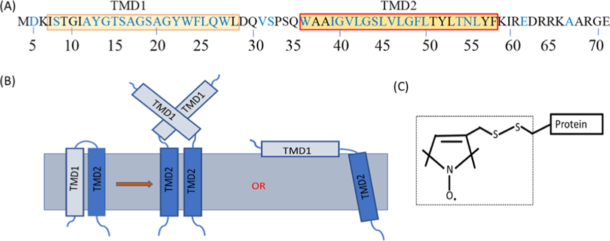
Figure 1.

(A) Primary sequence of S2168, boxes indicate TMD1 and TMD2. The amino acid positions studied by EPR spectroscopy are shown in blue. (B) Predicted topology of S2168 is adapted from the literature.1,2,12,38 TMD1 completely externalizes from the lipid bilayer and remains in the periplasm or partially externalizes and stays on the surface of the lipid bilayer, where TMD2 remains in the lipid bilayer. (C) R1 side chain shown in the dotted box which is attached to the protein through the disulfide bond of a Cys residue.
