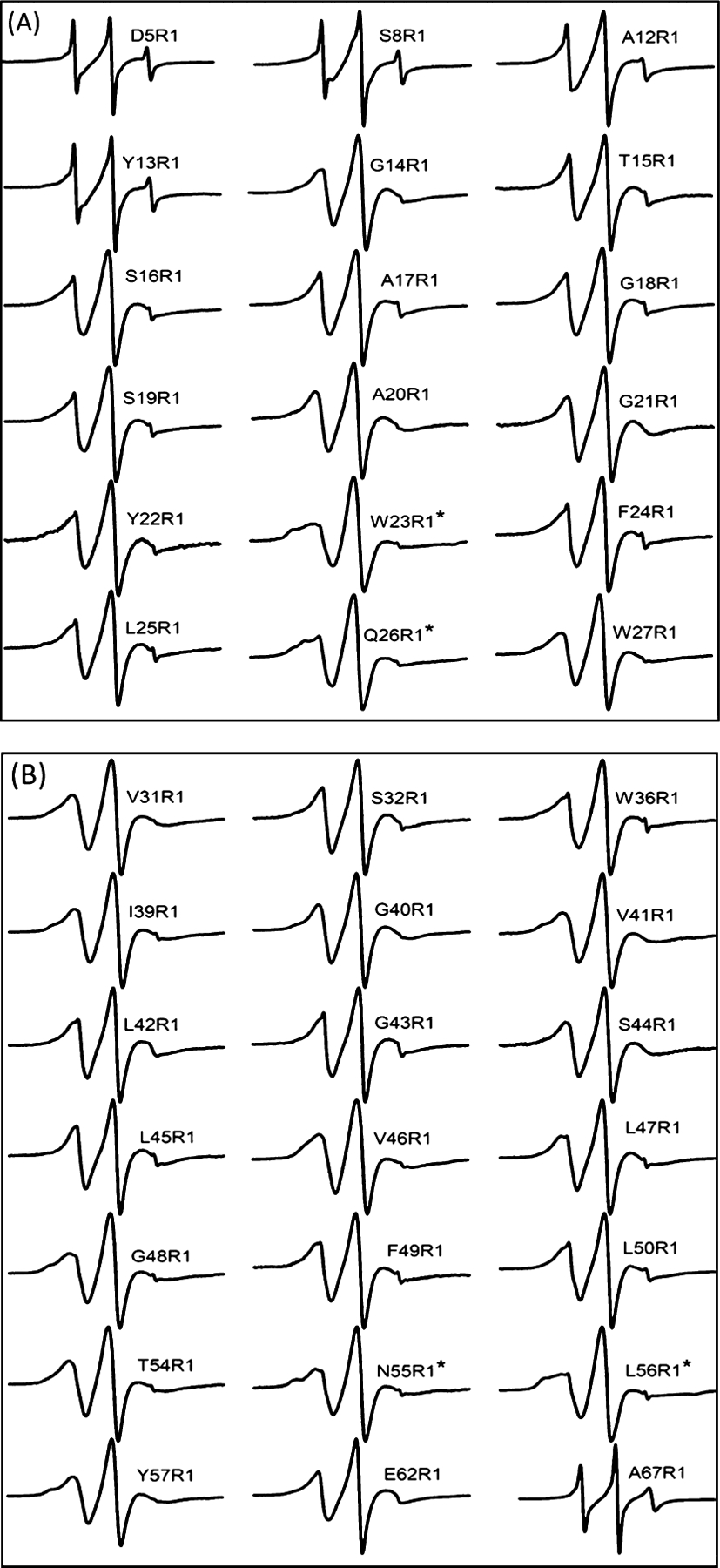
Figure 3.

CW-EPR spectra of R1 side chains attached at indicated positions by replacing the native amino acid with Cys. All spectra were normalized to the highest spectral intensity. (A) EPR spectra from the N-terminal and TMD1 of pinholin S2168. (B) EPR spectra from the loop to the C-terminal of pinholin S2168 including TMD2. CW-EPR spectra composed of multiple components were marked with (*).
