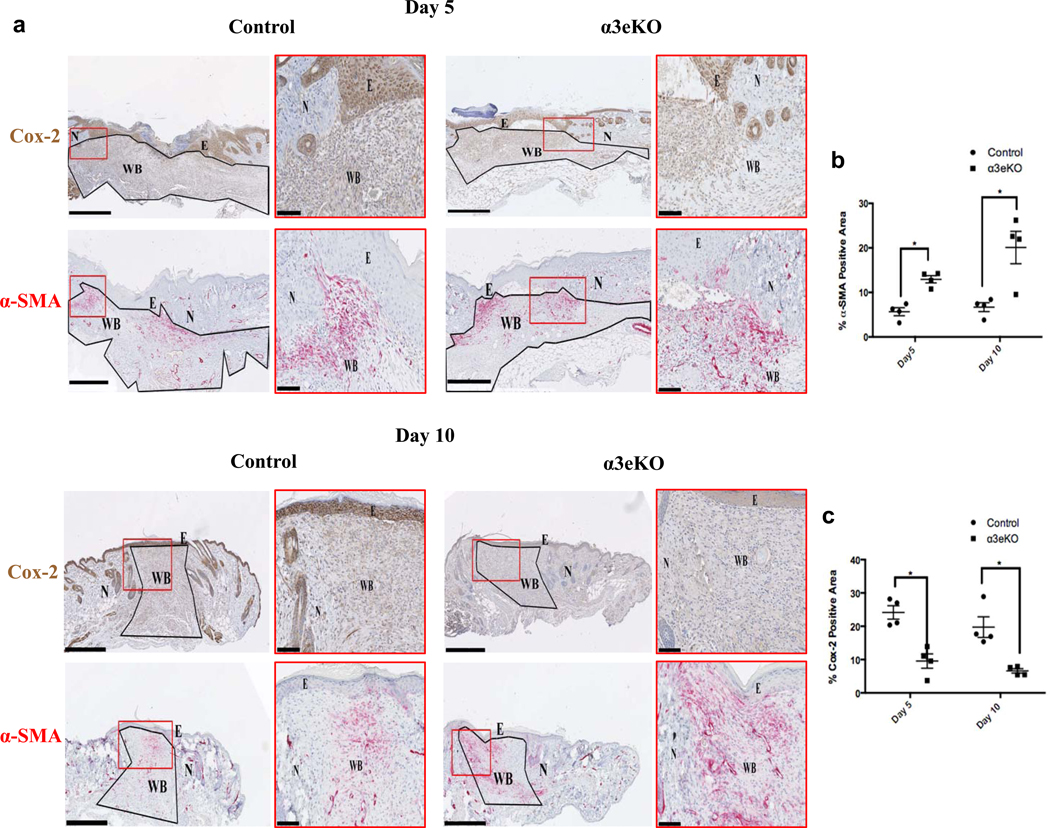
Figure 1. Genetic deletion of α3 in the epidermis leads to reduced Cox-2 expression and enhanced α-SMA expression in dermal cells of cutaneous wounds, in vivo.
(a) Mouse wound sections (5 and 10 days post wounding) from control and α3eKO mice were immunostained for Cox-2 (brown) or α-SMA (red). E=epidermis; N=normal skin; WB=wound bed; scale bar=500μm. Wound beds were outlined (black) and defined as the ROI. Insets (red boxes) illustrate staining and wound features (scale bar=100μm). Stains were separated by a color-deconvolution algorithm, and percentages of α-SMA-positive staining (b) or Cox-2-positive staining (c) within each ROI were quantitated (ImageJ). 2-way ANOVA followed by Bonferroni post-hoc analysis, *p > 0.05, n= 4 mice per time point / genotype.

