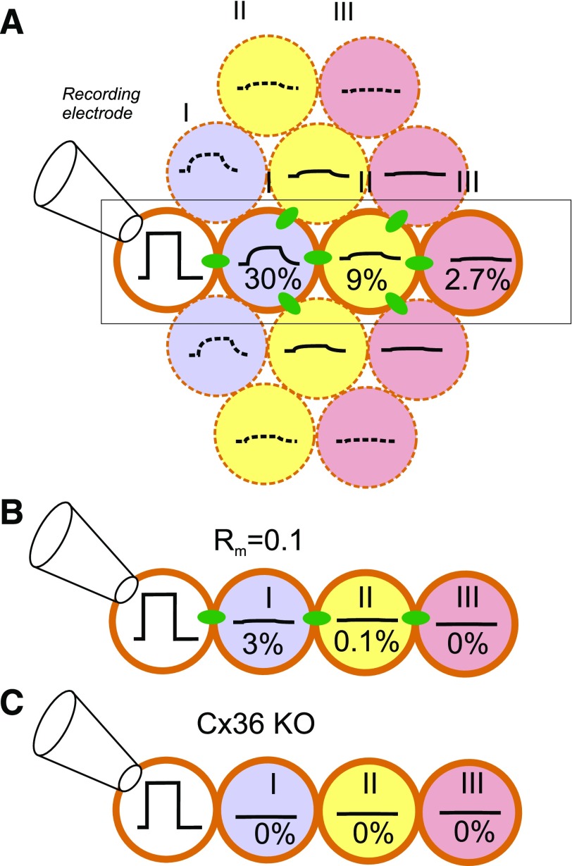
Figure 3.
Attenuation of electrical signaling via gap junctions. A: A voltage step is applied to a voltage-clamped β-cell in an islet (white) (connected to recording electrode). The change in membrane potential enters neighboring cells (blue) (layer II) via the gap junctions but is attenuated by 70%, and its time course is “filtered” by the resistive-capacitive properties of the β-cell. The voltage change in layer I extends into layer II (yellow) but undergoes additional attenuation, and only 9% remains. In layer III (pink), the signal has declined to <3% of the original value. B: Impact of increased KATP channel activity in a cell within layer I (due to, for example, expression of a gain-of-function mutant KATP channel or impaired ATP production). If the input resistance in layer I is reduced by 90%, then the voltage change in this and cells in layers II and III will be correspondingly smaller. C: Genetic ablation of connexin-36 (Cx36 KO) (the β-cell gap junctions) will abolish electrical coupling. B and C highlight the part of A indicated by rectangle.

