Figure 3.
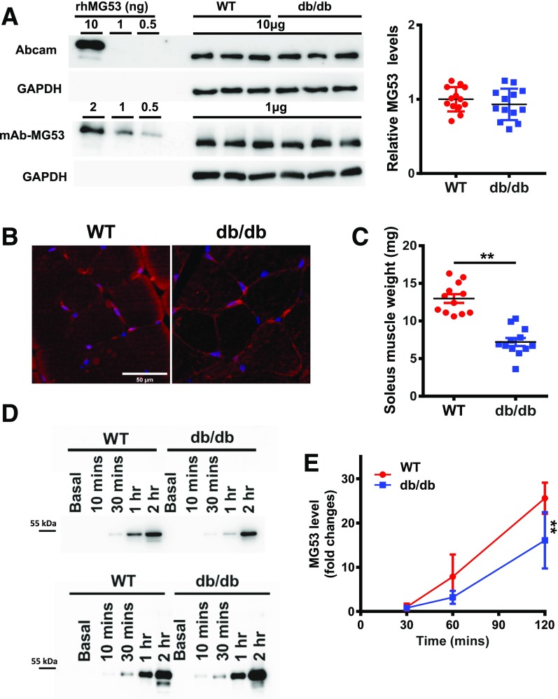
Muscle atrophy in db/db mice leads to reduced secretory activity of MG53. A: Skeletal muscles from WT and db/db littermate mice were probed with Abcam antibody (top) and mAb-MG53 (bottom). On average, there was no statistical difference in MG53 protein between WT and db/db muscle. B: Cross section of skeletal muscle derived from WT (left) and db/db (right) mice was stained with mAb-MG53. C: Comparison of soleus muscle mass derived from littermates of WT and db/db mice at 4 months of age (**P < 0.01). D: 20 µL out of a total of 2 mL solution bathing the soleus was loaded onto the gel at different time points after insulin treatment (top). Smaller amounts of MG53 were detected from db/db soleus at all time points. The bottom panel shows loading of the bathing solution normalized to soleus muscle mass. E: Time-dependent secretion of MG53 from WT (red) or db/db (blue) soleus muscle following insulin treatment (**P < 0.01).

