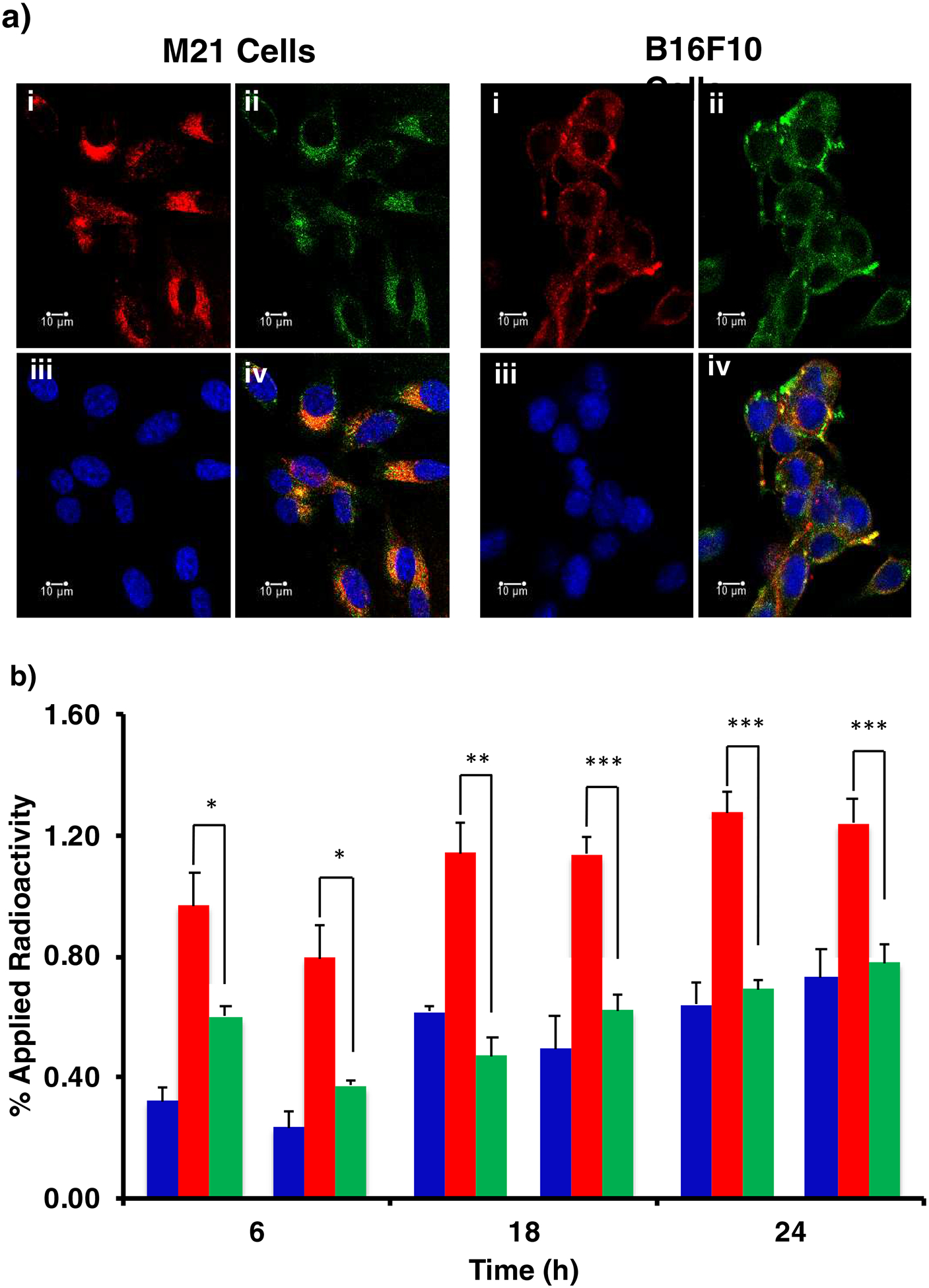
Fig. 3.

(a) Confocal fluorescence microscopy visualization of DOTA-αMSH-PEG-Cy5-C’ dots internalization in M21 human melanoma and B16F10 murine melanoma cells at 4 h post administration. M21 and B16F10 melanoma cells were imaged in the presence of DOTA-αMSH-PEG-Cy5-C’ dots (i), LysoTracker Green (ii) and Hoechst nuclei stain (iii). Merged images displaying all three probes (iv). Scale bar equals 10 μm. (b) Cell binding and internalization of 177Lu-DOTA-αMSH-PEG-Cy5-C’ dots in B16F10 and M21 melanoma cells. Membrane bound (◼) and internalized (◼) radioactivity were determined at 6 h, 18 h, and 24 h post incubation with 177Lu-DOTA-αMSH-PEG-Cy5-C’ dots. Co-incubation of 177Lu-DOTA-αMSH-PEG-Cy5-C’ dots with the MC1-R avid NDP blocking peptide (◼) was used to assess specificity of internalization. The significance of internalized versus blocked activity is given as *p<0.05. **p<0.01 and ***p<0.005. Each data point represents the mean ± s.d. of 3 replicates.
