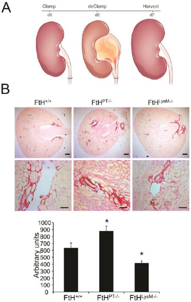
Figure 3. Role of myeloid-and proximal tubule-specific Ferritin heavy chain deletion on fibrosis during reversible obstructive nephropathy.

(A) Illustration of experimental design. Reversible obstructive nephropathy was induced by clamping one of the ureters for two days, following which the clamp was removed and animals were allowed to recover for five days. (B) Fibrosis following injury was determined by picrosirius staining on the obstructed kidney sections from wild-type (FtH+/+), proximal tubule specific FtH deletion mice (FtHPT−/−) and myeloid specific FtH deletion mice (ftHLysM−/−). Representative images of the stained kidney sections are shown in the upper panel (scale bar – 400 μm) and middle panel (scale bar – 100 μm). Lower panel: graphical representation of the collagen deposition in the kidneys. *p<0.05 vs FtH+/+ mice; n=5–6 per group. Reproduced with permission.10
