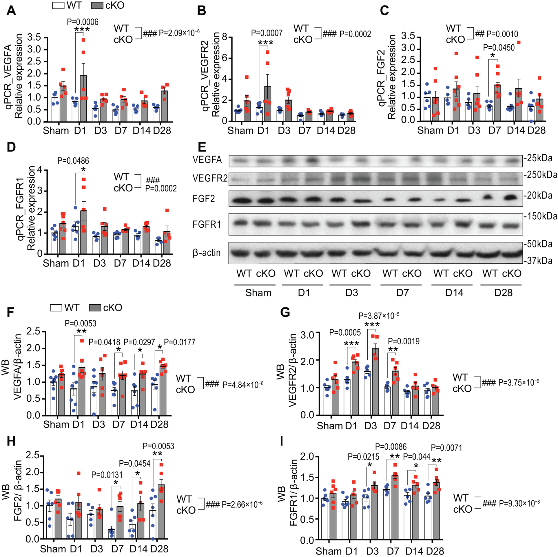
Fig. 6. Endothelium-targeted miR-15a/16–1 deletion enhances the expression of pro-angiogenic factors in ischemic mouse brains.

Total RNAs and proteins were isolated from the ipsilateral cortex of mouse brains at 1–28 d reperfusion after MCAO. qPCR and Western Blotting (WB) were carried out to detect the expression of pro-angiogenic factors. A-D, qPCR data showed elevated mRNA expression of VEGFA (A), VEGFR2 (B), FGF2 (C) and FGFR1 (D) in the cerebral cortex of EC-miR-15a/16–1 cKO mice than WT controls at two or more reperfusion time points after 1h MCAO. Accordingly, representative WB images (E) and quantitative analysis (F-I) indicated enhanced protein levels of VEGFA (F), VEGFR2 (G), FGF2 (H) and FGFR1 (I) in the cerebral cortex of EC-miR-15a/16–1 cKO mice than WT controls at three or more reperfusion time points after 1h MCAO. n = 5–7/group for all qPCR and WB experiments; *p < 0.05, **p < 0.01, ***p < 0.001 versus WT controls at each time point; ##p < 0.01, ###p < 0.001 for the overall difference between WT and EC-miR-15a/16–1 cKO groups; statistical analyses were performed by two-way ANOVA followed by Bonferroni’s multiple comparison tests.
