
Fig. 3.
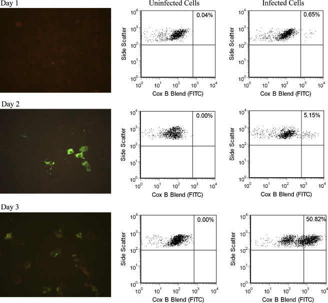
Comparison of IFA (left column) and flow cytometry analysis of uninfected PMK cells (center column) and PMK cells days 1–3 after inoculation with coxsackievirus B1 (right column). IFA showed detectable fluorescent cells with apple green cytoplasmic staining at day 2, while flow cytometry was able to detect a small number of cells with bright staining on day 1. Negative cells, counterstained with Evans Blue, are dull red on IFA.