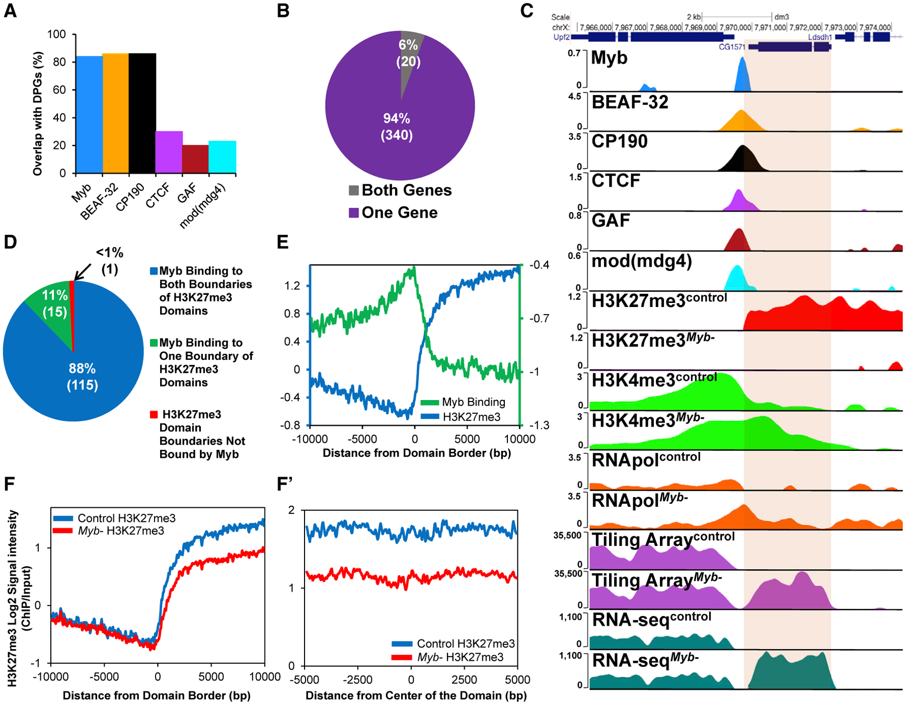
Figure 2. Myb Contributes to Insulator Function at DPGs and Binds to Boundaries of H3K27me3 PcG Domains to Maintain Domain Stability.

(A) Binding of Myb and insulator proteins to promoter regions of DPGs. Myb binds to 84% of all DGPs present in the genome (p = 2.32 × 10−275 compared to shuffled control), comparable to BEAF-32 and CP190 (~86% for both).
(B) Absence of Myb leads to changes in expression of genes associated with 360 DPGs.
(C) Example of upregulation of expression in one gene of a DPG in Myb mutants normally bound by Myb and insulator proteins. Loss of Myb leads to the reduction of H3K27me3, leading to the spreading of H3K4me3 and higher RNA pol II occupancy, with upregulation of CG1571, as shown with tiling microarray data, gene expression microarray data (153-fold upregulation, p = 5.77 × 10⁻²⁶), and RNA-seq data (397-fold upregulation, p < 0.0001).
(D) Myb is present at boundaries of 99% of all H3K27me3 domains previously described in D. melanogaster (p = 1.16 × 10⁻³² compared to shuffled control), with Myb binding to both boundaries of a domain 92% of the time.
(E–F′) Myb binding signal (purple line) is increased at boundaries of H3K27me3 domains (E; green line). Absence of Myb leads to reduced average levels of H3K27me3 at domain boundaries (F; red line) and continues across the length of the domains (F′; red line) (p < 0.0001, Student’s t test). DE, differentially expressed. See STAR Methods for sources of binding data.
