
Fig. 2.
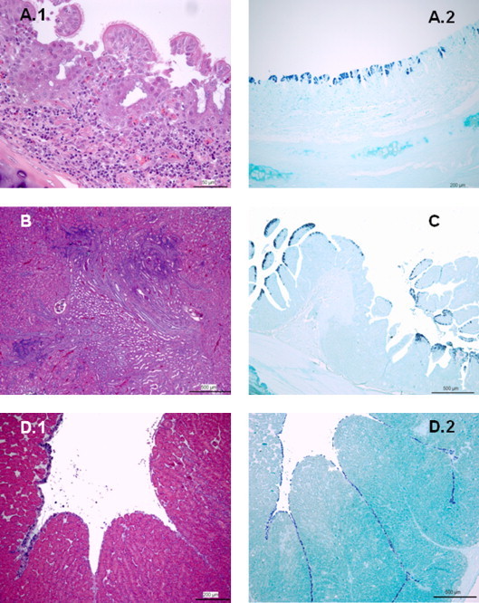
Adult hens infected with IBV Italy 02 serotype. (A) Trachea, day 2 PI. A.1: severe diffuse lymphocytic infiltration and hyperemia of lamina propia; note loss of glandular structures, mild hyperplasia and desquamation of ciliated epithelial cells lining tracheal mucosa (H/E; 50 μm); A.2: numerous ciliated epithelial cells of tracheal mucosa showed strong IBV genome specific staining in the cytoplasm (ISH; 200 μm). (B) Kidney, day 11 PI. Mild multifocal lymphocytic infiltration is observed in renal interstitium (H/E; 500 μm). (C) Rectum, day 2 PI. Numerous strong positive-stained enterocytes were observed in the rectal mucosa (ISH; 500 μm). (D) Oviduct, day 19 PI. D.1: desquamation and degenerative changes are observed in epithelial cells lining glandular tissue (H/E; 200 μm); D.2: extensive IBV genome specific staining of epithelial cells is shown (ISH; 500 μm).