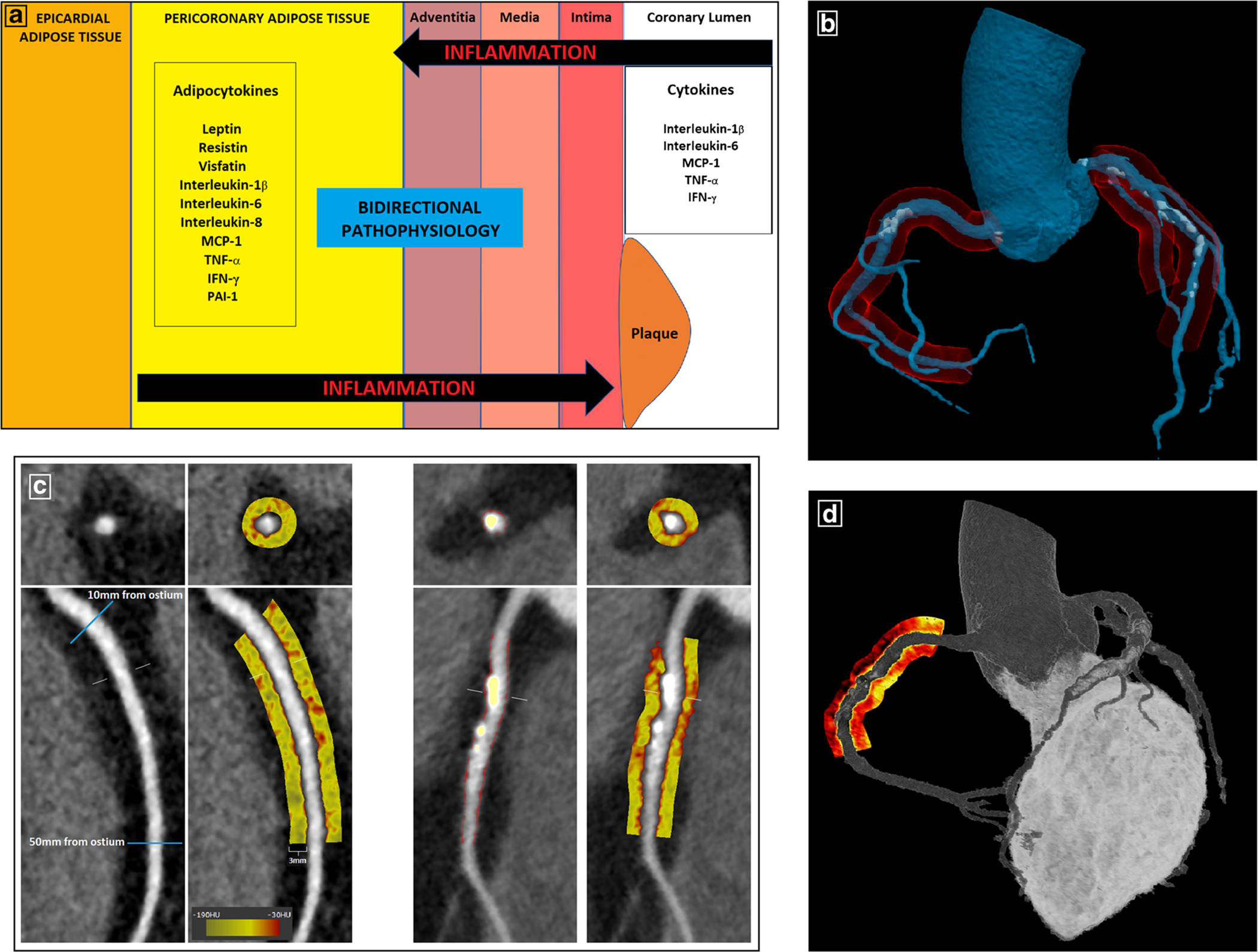
Fig. 1.

Pericoronary adipose tissue—from biology to imaging phenotyping. a Bidirectional communication between PCAT and the coronary arterial wall. Dysfunctional PCAT secretes pro-inflammatory adipocytokines which diffuse directly into the vessel wall and contribute to atherosclerosis via paracrine and vasocrine mechanisms. The recent discovery of “inside-to-outside” signaling pathways demonstrates that PCAT can also function as a sensor of coronary inflammation. b Schematic representation of PCAT (red) surrounding the 3 major coronary arteries in a 3D anatomical model generated from CCTA. c PCAT quantification on CCTA using semi-automated software (Autoplaque v2.5). Left-sided panels show the proximal segment of the RCA (10–50 mm from RCA ostium) in curved and cross-sectional views, with PCAT visualized within a 3-mm radius around the vessel on a color map (Hounsfield unit scale inset). Right-sided panels show plaque quantification in the proximal RCA (non-calcified plaque in red overlay and calcified in yellow overlay) and the corresponding PCAT color map. d 3D rendering of a PCAT “heat map” around the proximal RCA
