
Figure 6.
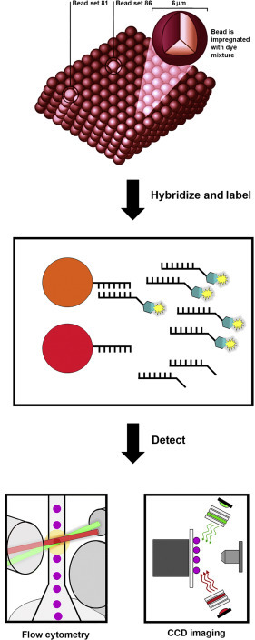
Suspension bead array. Microspheres (beads) are internally dyed with different intensities of red and infrared dyes to create 500 beads sets with unique spectral identities. Bead sets coupled with specific oligonucleotide probes are hybridised to amplified targets generated from the starting sample. Hybridised targets are labelled using a fluorescent reporter dye, and the microsphere suspension is analysed by flow cytometry or CCD imaging to identity each bead and quantify the reporter probe-target reaction on the microsphere surface.