
Fig. 4.
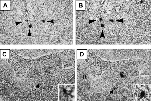
Identification of cells supporting PRRSV replication in secondary lymphoid organs during asymptomatic infection. Photomicrographs, taken at low magnification, showing in situ hybridization results in adjacent thin sections of a lymph node (A and B) at 63 dpf and a tonsil (C and D) at 132 dpf. The arrows identify RNA-positive cells that are present in both sections. Inset is a higher magnification showing the hybridization signal.