
Figure 4.
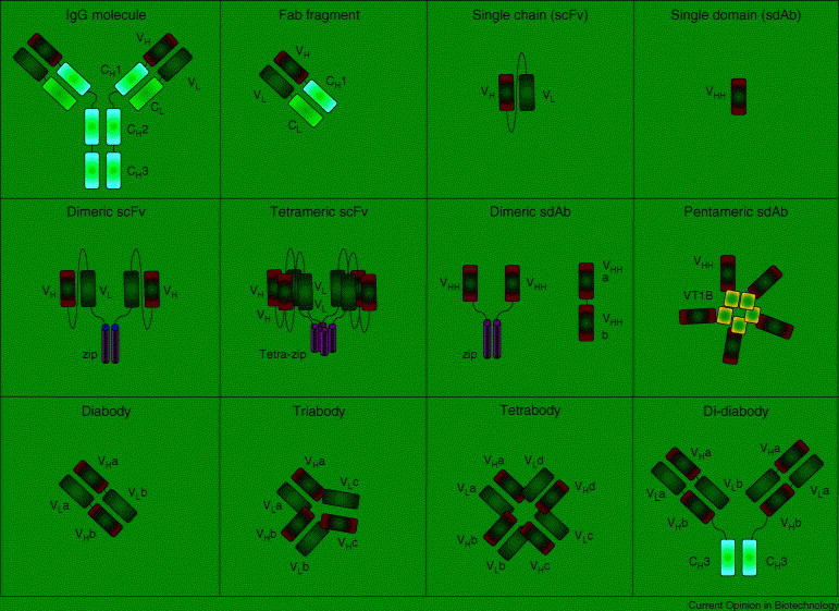
Structures of common recombinant antibodies. Schematic drawing showing the domain structure of various rAbs (Fabs, scFvs and sdAbs) and some of their oligomeric formats discussed in the text. The structure of a natural IgG molecule is also given for reference. Constant domains (C) are in blue and variable domains (V) in yellow; Ig domains are depicted as rectangles for both the heavy (H) and light (L) chains. Amphipathic α helices with dimerization (zip) and tetramerization (tetra-zip) capacity are shown in red. The pentameric B subunit of Verotoxin 1 (VT1B) is shown as a green square.