
Fig 7.
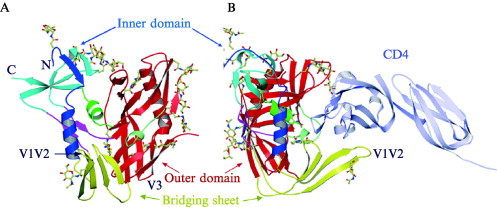
Structure of the gp120 core in the CD4‐bound state (Kwong et al., 1998). (A) Ribbon representation, showing the inner and outer domains, linked by a bridging sheet. The locations of the V1–V2 and V3 loops, deleted from the core construct (compare with Fig. 6), are shown. The locations of carbohydrate chains are shown by molecular ball‐and‐stick representations of those sugars found to be well‐ordered in the crystal structure. The view is into the CD4‐binding pocket. N and C termini of the core are labeled. (B) Side view of the same structure, with the first two immunoglobulin‐like domains of CD4 also shown.