
Fig 9.
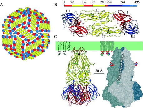
The flavivirus fusion protein E. (A) Organization of E dimers in the virion surface. Each subunit is shown in three colors: domain I in red, domain II in yellow, and domain III in blue—based on the structure described by Kuhn et al., (2002). (B) The soluble ectodomain, sE of dengue virus type 2, in the dimeric prefusion conformation found on mature virions (Modis et al., 2003). The domain colors are as in (A). The bar above the ribbon diagram shows the relationship of domains to primary structure. The “stem” segment between residue 394 and the transmembrane anchor is not included in the three‐dimensional structure. (C) The sE trimer (Modis et al., 2004). The proteins are shown in relation to a schematic lipid bilayer to illustrate the likely degree of penetration of the fusion loops (top) into the membrane. The ribbon diagram (left) is colored as in (A) and (B). Arrows at the C terminus of the polypeptide chain suggest its presumed continuing direction. The surface rendering (right) includes a dashed arrow to show the proposed course of the stem peptide, which would lead to the transmembrane anchor.