
Fig 10.
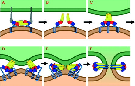
Diagram of membrane fusion by class II viral fusion proteins. (A) Receptor binding through domain III of E (flaviviruses). (B) Lowered pH in an endosome leads to dissociation of the dimer interactions. On release of dimer constraints, monomers can flex outward, presenting their fusion loops to the target cell membrane. (C) Insertion of the fusion loops into the target cell membrane and initial formation of trimer contacts among the projecting domains II. (D) Domain III flips over and the stem zips up along the outside of the trimer. (E) Hemifusion stalk. The diagram shows a proposed role for the inserted fusion loops—stabilization of the hemifusion stalk. (F) Formation of a fusion pore. Completing the zipping up of the stem drives fusion forward, because the cytosolic tails enter the pore and commit it to dilation.