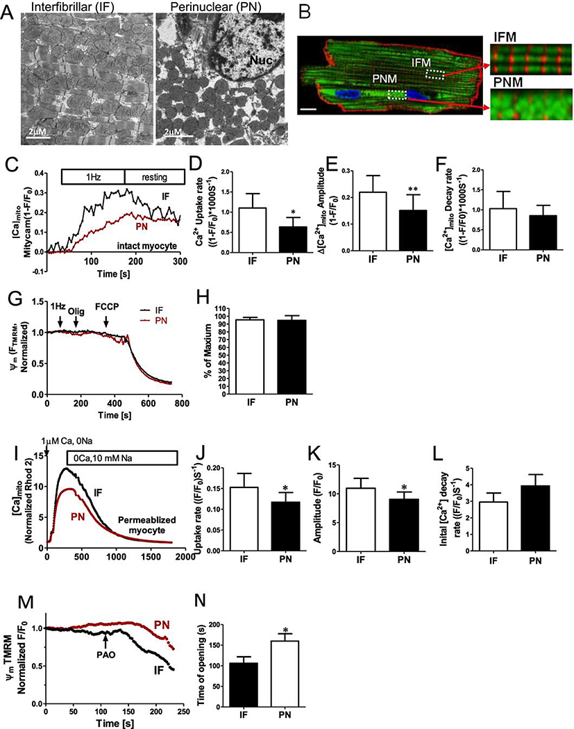
Figure 1. Perinuclear and intermyofibrillar mitochondrial morphology and Ca2+ uptake in intact cardiac myocytes.
(A) Electron microscopy images of sections through left ventricular rabbit heart. Intermyofibrillar mitochondria (IF) are often a rounded brick-shape (left), and perinuclear mitochondria (PN) are more round and loosely arranged (right). (B) Ad-Mitycam indicates mitochondria (and [Ca]mito), Di-8-ANEPPS indicates T-tubule (red) and Hoechst 33342 indicates the nucleus. Enlarged images from the indicated myocyte regions in B (scale bar 10 μm). (C) Kinetics of [Ca]mito during 1 Hz pacing frequency in adult rabbit cardiac myocytes, mean initial [Ca]mito uptake rate (D), Δ[Ca]mito amplitude (E) and decline rate[Ca]mito when pacing stopped (F; n=6 cells). (G,H) Mitochondrial membrane potential Δψm after 1 Hz pacing, normalized to the maximal value with oligomycin (n=6 cells). Kinetics of [Ca]mito change during Ca2+-clamp in permeablized (and SR disabled) cardiac myoyctes (I), mean Δ[Ca]mito (K), initial uptake rate (J) and [Ca]mito decline rate (L; n=6 cells). M,N ψm was normalized to the initial value, and mPTP opening time induced by phenylarsine oxide (PAO; n=4 cells) was estimated as the time when a regression line for the first 20 points of sustained ψm decay intersect the baseline.

