
Figure 1.
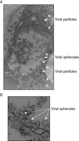
Representative electron micrographs of portions of N. benthamiana cells infected with the tombusvirus Cucumber necrosis virus (CNV). (A) The CNV-induced spherules in the center of the image and the assembled large number of virions in plant cells are depicted with arrows. Note that the entire cytosol of the portion of the cell shown is completely filled by CNV virions, demonstrating robust CNV replication. Magnification is 49,000×. (B) Several characteristic CNV-induced spherules are marked with arrowheads on the EM images. These 50–80 nm spherules are formed via membrane invagination into peroxisomal or ER-derived membranes. Narrow openings (necks) are visible likely connecting the spherules to the cytosol. Control samples lacking CNV do not show similar structures (not shown). Magnification is 98,000×. The images were taken by Dr. Barajas.