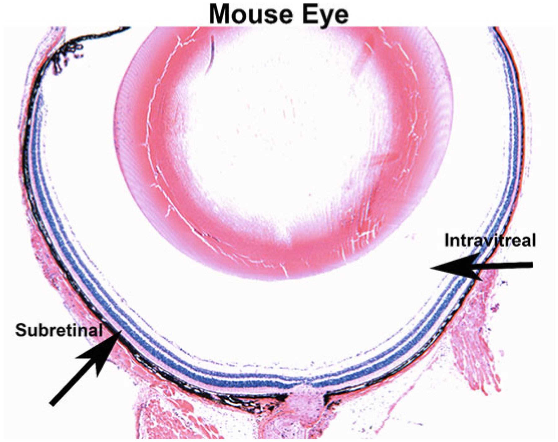
Fig. 4.

Subretinal and intravitreal injection sites in the mouse eye. A cross-section of a mouse eye stained with hematoxylin and eosin. Subretinal and intravitreal injection locations are marked by black arrows, where the subretinal injection creates a viral bleb between the photoreceptor cells and the retinal pigment epithelium (RPE) and the intravitreal injection places the virus into the vitreous fluid of the eye
